Introduction
MR Venography is a special imaging test that helps doctors see veins inside your body. This test uses magnetic resonance imaging (MRI) to create clear pictures of your veins. Because it is safe and does not use X-rays, MR Venography is important for finding blood clots or other vein problems. Many people need this test to help their doctors make the right diagnosis.
What is MR Venography?
MR Venography, also called MRV, is a type of MRI scan. It focuses on showing the veins, which are blood vessels that carry blood back to your heart. Unlike regular MRI, MR Venography highlights the veins so doctors can spot blockages, clots, or other issues. This test is often used when doctors need a clear view of the veins in your brain, legs, or other parts of your body.
Indications: When is MR Venography Needed?
Doctors may recommend MR Venography for several reasons. For example, it helps find blood clots, which can be dangerous if not treated. Sometimes, it is used to check for narrowing or blockages in the veins. In addition, MR Venography can help diagnose:
If you have symptoms like swelling, pain, or headaches, your doctor may suggest this test.
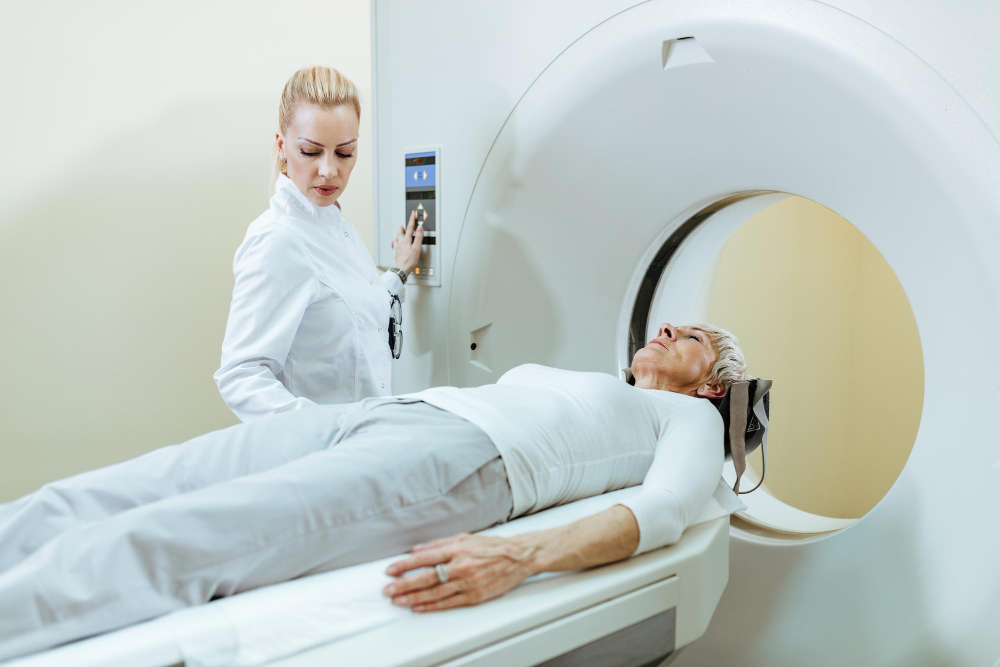
How MR Venography Works
MR Venography uses a strong magnet and radio waves to take pictures of your veins. First, you will lie on a table that slides into the MRI machine. Sometimes, a special dye called contrast is injected into your vein. This dye makes the veins show up more clearly on the images. The machine then takes detailed pictures, which doctors use to check for problems. Because there is no radiation, MR Venography is considered safe for most people.
Preparation: How to Get Ready for the Test
Getting ready for MR Venography is simple. However, you should follow these steps to make sure the test goes smoothly:
Because some people feel nervous in small spaces, let your doctor know if you are claustrophobic. They can help you feel more comfortable.
What to Expect During the Procedure
When you arrive, a technologist will explain the steps. Next, you will lie on a table that moves into the MRI machine. If contrast dye is needed, it will be given through a small needle in your arm. During the scan, you will hear loud noises, but earplugs or headphones are usually provided. The test usually takes 30 to 60 minutes. While the scan is happening, you must stay very still so the images are clear. After the test, you can go home and return to normal activities unless your doctor tells you otherwise.
Risks and Safety Information
MR Venography is safe for most people. Because it does not use radiation, there is less risk than with some other scans. However, there are a few things to keep in mind:
According to the World Health Organization and the Centers for Disease Control and Prevention, MR Venography is generally safe when performed by trained staff.
Results: Understanding Your MR Venography Report
After the test, a radiologist will review your images and write a report. This report will explain if there are any blood clots, blockages, or other vein problems. Your doctor will discuss the results with you and explain what they mean for your health. If treatment is needed, your doctor will guide you on the next steps.
Frequently Asked Questions
Conclusion and Call-to-Action
MR Venography is a safe and effective way to check for vein problems. Because it uses advanced imaging, doctors can find issues early and plan the right treatment. If you have questions or need more information, consult a radiologist at Shreeji MRI or your healthcare provider for personalized guidance about MR Venography.